
Tools
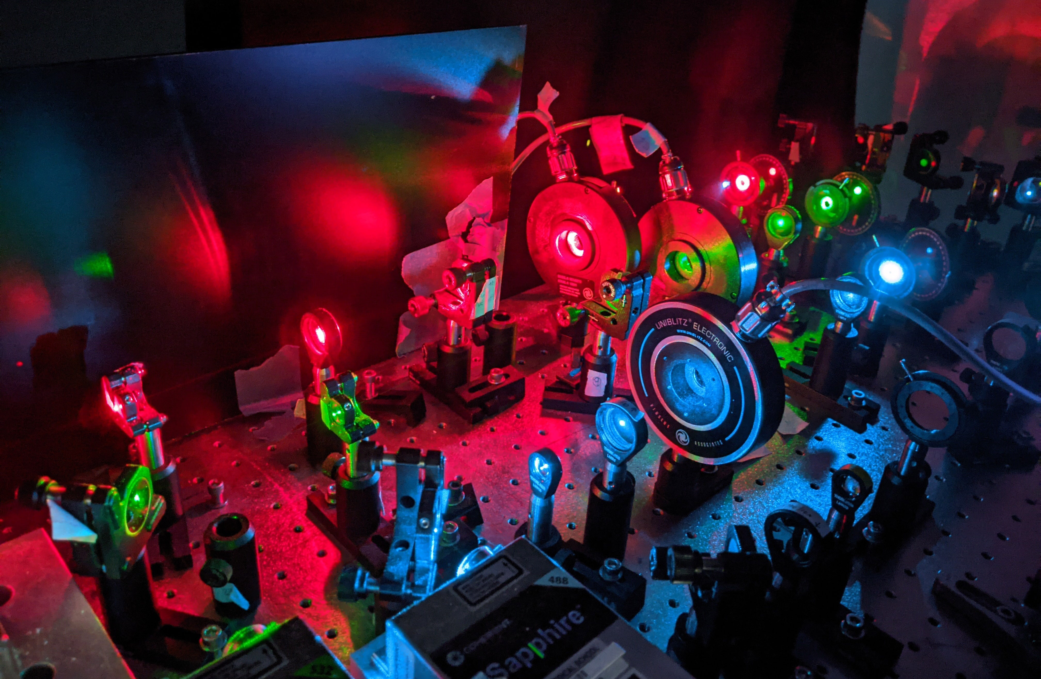
We use a broad range of single-molecule approaches in vitro and in cells. We also enjoy building microscopes. Explore this page to learn more about what we are up to! We are particularly interested in developing new single-molecule assays to better probe the structure and function of biological machines. Examples from our published work include methods that investigate:
- How DNA binding proteins compact DNA
- How polymerases exchange at DNA lesions in vitro and in cells
- How the NHEJ synaptic complex forms and how its enzymatic activities are coordinated with synapsis
- How mechanical force influences protein function
- How structural dynamics are coupled to enzyme function
The Harvard Medical School News Office made a video series describing some of our microscopes and how we name them.
Read the full story at https://hms.harvard.edu/news/action-heroes-dna-repair
Watch the 6-part video series at https://www.youtube.com/watch?v=lwtE3dkwIpM&list=PL9fwcf7k-_bHj90CmL_RjY05BoCKeLyu-
In a single molecule Forster resonance energy transfer (smFRET) assay one measures the efficiency of energy transfer between a donor and acceptor fluorophore. This energy transfer is highly sensitive to the distance between the two dyes and occurs over a range of distances (~30-90 Angstroms) that is particularly relevant for biological molecules. We have used FRET extensively to monitor the distance between DNA ends during non-homologous end joining(NHEJ), a prominent DNA double strand break repair pathway (Graham et al. 2016). Modifications of this assay have allowed us to probe protein binding dynamics and stiochiometry (Graham et al. 2018) or enzymatic activity of end processing factors (Stinson et al. 2020) and correlate this with the structure of the NHEJ synaptic complex. We have also used smFRET approaches to study conformational dynamics of proteins (Catapovic et al. 2019). Currently we are developing three-color smFRET assays to study the conformational dynamics of NHEJ factors within the synaptic complex. The movie shows how changes in FRET efficiency due to formation of the short-range synaptic complex are correlated with changes in the occupancy of the core NHEJ factor XLF (the blue signal).
We have developed novel single-molecule assays to better track the motion of flow stretched DNAs and to detect the binding and action of unlabeled DNA binding proteins. In these approaches double stranded DNA is attached to the surface of a microfluidic flow cell and extended under buffer flow. Hydrodynamic drag along the DNA results in tension varying across the DNA molecule; tension is highest at the tether point while it approaches zero at the free DNA end. We have site-specifically labeled DNA with quantum dots and visualized the motion of distinct segments of DNA, an approach we named DNA motion capture (Price et al. 2015). Given the flow-induced force titration along DNA, we showed that one can distinguish distinct modes of DNA compaction by distinguishing how different DNA segments are compacted by DNA binding proteins (Graham et al. 2014). Furthermore, we showed how protein induced fluorescent enhancement (PIFE) can be used to detect proteins binding to DNA and inducing DNA compaction (see movie on left and Song et al. 2016). We have used these approaches broadly to understand how DNA binding proteins condense DNA.
UNDER CONSTRUCTION...
In single-particle tracking Photoactivation Localization Microscopy (sptPALM), individual fluorescent proteins are photoactivated and then imaged until they photobleach. sptPALM enables one to follow the dynamics of individual proteins at high concentrations in living cells. We have been using sptPALM in E coli where we image fluorescent protein fusions to components of the DNA replication machinery and low fidelity translesion polymerases. By tracking the motion of individual polymerases in cells upon treatment with DNA damaging agents, we have investigated how translesion polymerases are recruited to the replisome and defined the critical interactions in this multistep process (see Thrall et al 2017 and Chang et al. bioRxiv 2020). This movie (see left) shows the activation an imaging of a epsilon, a subunit of the replicative polymerase Pol III.
UNDER CONSTRUCTION... We apply force to biological molecules in order to (1) probe how force impacts protein structure and function and (2) to distinguish reaction intermediates by their elastic properties.